FTMap: fast and free* druggable hotspot prediction

*free to academics

FTMap is a useful and fast online tool that attempts to mimic experimental fragment-screening methodologies (SAR-by-NMR and X-ray crystallography) using in silico methods.   The algorithm is based on the premise that ligand binding sites in proteins often show “hotspots” that contribute most of the free energy to binding.

Often, fragment screening will identify these hotspots when clusters of different types of fragments all bind to the same subsite of a larger binding site.   In fact, x-ray crystallography studies of protein structures solved in a variety of organic solvents demonstrate that small organic fragments often form clusters in active sites.

In the FTMap approach, small organic probes are used for an initial rigid-body docking against the entire protein surface.  The “FT” of FTMap stands for the use of fast Fourier transform (FFT) methods to quickly sample billions of probe positions while calculating accurate energies based on a robust energy expression.

Following docking of each probe, thousands of poses are energy-minimized and clustered based on proximity.  The clusters are then ranked for lowest energy.   Consensus sites (“hot spots”) are determined by looking for overlapping clusters of different types of probes within several angstroms of each other.   If several consensus sites appear near each other on the protein surface, that is a strong indication of a potentially druggable binding site.

Isotopic labeling of proteins in non-bacterial expression systems

As therapeutic proteins gain importance alongside traditional small molecule drugs, there is increasing interest in using NMR methods to examine their structure, dynamics, and stability/aggregation in solution.

Modern heteronuclear NMR of proteins relies on isotopically-labeled samples containing NMR active nuclei in the peptide backbone, sidechains, or both.

Although isotopic-labeling of recombinant protein is typically carried out in E. Coli expression systems, many biotherapeutic proteins must be expressed in eukaryotic systems to insure proper folding and/or post-translational modifications.   In practice, this means overexpression in either yeast, insect or mammalian cells.

Increased interest in attaining labeled protein samples for analysis by NMR is leading to better commercial availability of isotopically-labeled expression media and improved vectors for overexpression in non-bacterial systems.

Comprehensive reviews of state-of-the-art protocols and procedures for expression of isotopically-labeled proteins in non-standard systems are available here: yeast, insect cells, and mammalian cells.


Virtual screening capability for under $5K?

Many early stage companies may be missing out on the value that docking can provide at the validated hit and hit-to-lead stages of development, where structure/activity relationships (SAR) can help guide chemistry development of lead compounds.

While docking large HTS libraries with millions of compounds may require specialized CPU clusters, docking of small libraries (i.e., thousands of compounds) and SAR compounds from experimental assays is readily achievable in short time frames with a relatively inexpensive Intel Xeon workstation.

Following an initial investment in the workstation and software, follow-on costs are minimal (e.g., electricity, IT support and data backup). Turnaround times may be faster than with CRO services.  Also, sensitive IP data is also protected by being retained onsite and not transmitted over the internet.

Equipment / cost breakdown:

Software:

AutoDock Vina (non-restrictive commercial license)       cost: free

Accurate (benchmarked against 6 other commercial docking programs)

Compatible with AutoDock tools

Optimized for speed (orders of magnitude faster than previous generation)

Parallelized code for multi-core systems

AutoDock Tools (non-restrictive commercial license)      cost: free

PyMol Incentive (commercial license)                                        cost ~$90 / mo

Visualize docking results, free plugin can allow Vina to be run within PyMol GUI

Fedora Linux                                                                                              cost: free

Hardware:

HP Z620 Workstation (stock configuration)                          cost: $2999

2 GHz (6 Core) Intel Xeon E5-2620 2GHz

USB keyboard and mouse                                                                  cost: $50

Dell Ultrasharp 27” LED monitor                                                  cost: $649

1TB USB HD for data backup                                                          cost: $150

IT support for initial setup ~ 4 hours                                           cost: $400

Total initial capital expenditure:                                                  ~$4350


10 Common Mistakes in Fragment Screening

There is an excellent review paper from Dan Erlanson and Ben Davis that came out last year detailing some of the more common mistakes and artifacts that can arise in fragment-based screening campaigns (so-called “unknown knowns”).  I encourage readers to go read the original paper.  I have summarized some of the key points below:

1) Not checking compound identity to make sure what you think you purchased is what you actually have.

2) Low-level impurities in compound stocks can cause problems at the high concentrations used in fragment screens.

3) DMSO, commonly used to store fragments in plates, can act as a mild oxidant and is also hygroscopic.

4) Pan-assay interference compounds (PAINS) are common in many libraries and are found to give false positives to many targets.

5) Reactive functional groups in fragment hits can cause covalent binding or aggregation of the target.

6) Many fragments can show binding or inhibition while acting as aggregators rather than reversible binders.  Including a small % of detergent can help eliminate these kinds of fragments from giving positive signals.

7) STD-NMR is very sensitive to weak binders, but because it relies on a relatively fast disassociation rate for the ligand, tight binders (<1 uM) can be missed by this method.

8) X-ray crystallographic structures are often taken as the “truth” when they are in fact a model of an electron density.  Fragments can often be modeled into the density in incorrect orientations or in place of solvent atoms.

9) SPR methods are very sensitive to fragment binding, but can be confounded by non-specific binding of fragment to the target or chip, as well as compound-dependent aggregation.

10) Fragment hits should be validated by more than one method before embarking on optimization.  They should also be screened for being aggregators by DLS or other methods.

Gilead’s innovative approach to Hep C drug, Sovaldi

Hepatitis C virus (HCV) is a single-stranded RNA virus that infects an estimated 180 million people worldwide.

In 2013, Gilead received FDA approval for a new HCV drug, Sovaldi (sofosbuvir), that inhibits viral replication by targeting the virus’s NS5B polymerase.  Sovaldi has shown a very high cure rate (nearly 100% HCV suppression and sustained virological response) in clinical trials of previously untreated patients and has fewer side effects than pegylated-interferon and ribavirin therapies.

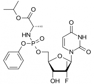
Sovaldi is a methyluridine-monophosphate prodrug: it is metabolized in the body back into methyluridine-triphosphate, which acts as a potent substrate mimic and inhibitor of the NS5B polymerase.

What is interesting about Sovaldi is the approach the scientists took to getting the inhibitor into the cell, relying on phosphoramidate prodrug  technology that had been effectively used to develop anti-HIV drugs, but had never been applied before to this class of anti-HCV drugs.

During development, the researchers decided that they needed to deliver the charged methyluridine-monophosphate (rather than the neutral methyluridine)  into the cell on the basis of two key observations:  1) the methyluridine triphosphate is the active compound against HCV NS5B polymerase, while the methyluridine alone is inactive (owing  to very low conversion to monophosphate in vivo) and 2) the methyuridine monophosphated derivative can be anabolized in the cell back to the potent triphosphate form by an endogenous uridine-cytidine monophosphate kinase.

jm-2010-00863x_0003
The active uridine triphosphate (6) can be created when 4 is metabolized to methyluridine-5′-monophosphate. Compound 5 is not phosphorylated and is inactive in cells.


The phosphoramidate prodrug technology had never been applied to HCV inhibition until Solvadi.

The idea behind  phosphoramidate prodrug technology is to create a membrane-soluble neutral prodrug derivative that can be metabolized in the liver by carboxylesterase-mediated cleavage and subsequent steps back to the monophosphate form.

The researchers applied the approach and after a significant amount of  SAR investigation and PK/PD studies around the chemical composition of the phosphoramide substituents, they concluded that the structure of compound shown above was the optimal structure to deliver the methyluridine-monophosphate to the liver.

The result is a new generation of highly effective HCV therapeutics with few side effects that can make a significant difference in the lives of patients living with HCV.